
Breast cancer continues to be a significant global health challenge, and accurate screening methods are crucial in its battle. While mammography has long been the standard for breast cancer screening, its limitations are becoming increasingly evident, particularly in individuals with dense breast tissue. Dense breast tissue not only heightens the risk of breast cancer but also makes tumor detection considerably more challenging as it can obscure anomalies on mammograms. In this context, an innovative development has emerged from Microsoft’s AI for Good Lab, led by Felipe Oviedo, Ph.D. By harnessing the power of artificial intelligence, this new model significantly enhances the accuracy of detecting breast cancer through MRI, promising to revolutionize screening practices and address the shortcomings of traditional methods.
Bridging Screening Gaps with AI Technology
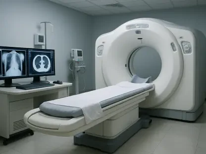
Current screening methods primarily rely on mammography, which, despite being widely adopted, has limitations, especially with dense breast tissue. Studies show that breast MRI offers higher sensitivity and can serve as a supplemental tool. However, the high costs and elevated false-positive rates associated with MRI pose significant challenges to its broader adoption. Addressing this complexity, researchers at the University of Washington, in partnership with clinical investigators, developed an AI model specifically designed to improve the efficiency and accuracy of MRI in breast cancer screening. Anomaly detection technology forms the core of this model, contrasting starkly with earlier binary classification models. These older models were often critiqued for their unrealistic data distributions, limiting their application in real-world settings.
The innovative AI model developed by Dr. Oviedo’s team utilizes a vast dataset of nearly 10,000 contrast-enhanced breast MRI exams amassed over 17 years. This formidable dataset predominantly featured white female patients, with a significant portion presenting dense breast issues. Such a comprehensive dataset allowed the AI model to fine-tune its ability to recognize benign cases while remaining sensitive to malignant anomalies. This capability makes it particularly adept at discerning potential cancerous lesions, even if they were underrepresented in the training data.
Advancing Radiological Precision with Heatmaps
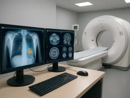
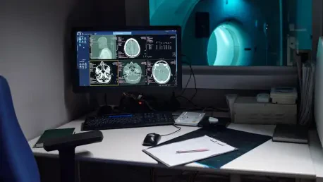
The model’s sophistication lies in its dual outputs: a precise anomaly score and spatially resolved heatmaps that visually highlight abnormal regions on MR images. This visual aid surpasses previous models by producing highly accurate depictions that align closely with areas of concern identified by radiologists. During rigorous testing phases, the AI model exhibited remarkable accuracy in identifying tumor locations. Both internal datasets from University of Washington exams and external datasets from a multicenter study confirmed its superior performance over benchmark models. Moreover, this model demonstrated consistent excellence across various testing scenarios concerning cancer prevalence detection tasks.
By integrating AI’s computational power, this model holds the potential to streamline radiological workflows, offering a triage system that distinguishes normal scans from those requiring focused reviews. This efficiency empowers radiologists to dedicate their expertise to cases with a higher likelihood of cancer, enhancing diagnostic precision and patient care. The anomaly detection and visual heatmaps provide a clear, interpretable guide, proving invaluable in clinical decision-making environments.
The Path Forward in Medical Imaging
Dr. Oviedo’s model not only introduces advancements in technology but also marks a step forward in personalized medicine, tailoring diagnostics more closely to individual risk factors. Integral to the model’s success is its transparency and ability to provide pixel-level explanations of abnormal regions. These capabilities cater to the growing need for models that radiologists can trust and apply confidently in practice. Moving forward, Dr. Oviedo stresses the necessity of further validation through larger datasets and prospective studies to ensure the model’s efficacy and reliability in diverse clinical settings.
The implications of this technology extend beyond simply detecting tumors. It aims to refine the approach toward cancer diagnostics by integrating cutting-edge technology with expert medical knowledge. Advancements like this could drastically redefine how breast cancer screenings are conducted, making them more sustainable and accessible while aligning with personalized diagnostic approaches. As the medical community pushes towards precision medicine, the role of AI becomes increasingly pivotal in shaping the future of healthcare.
Future Prospects for AI Integration
Current breast cancer screening primarily employs mammography, a method with limitations, especially for dense breast tissue. Mammography’s inadequacies have led to studies supporting MRI’s higher sensitivity as a supplemental tool, despite its high cost and frequent false positives hampering broader use. Researchers at the University of Washington, collaborating with clinical experts, created an AI model aimed at enhancing MRI’s efficiency and accuracy in breast cancer screening. The model is founded on anomaly detection technology, offering a sharp contrast to older binary models criticized for unrealistic data distributions, thus making them less practical in real-world scenarios.
Dr. Oviedo’s team pioneered an AI model using an extensive dataset of nearly 10,000 contrast-enhanced breast MRI exams collected over 17 years. The dataset mainly featured white female patients with significant dense breast tissue cases. This rich dataset enabled the AI model to excel at identifying benign cases while remaining keen to malignant ones, improving its ability to detect potentially cancerous lesions that might be rare in the training data.