
Nuclear cardiology has revolutionized cardiovascular diagnostics and treatment, providing detailed assessments of heart health with minimal invasiveness. The evolution of nuclear cardiology imaging systems has significantly improved our ability to detect and manage heart diseases, benefiting patients with higher diagnostic accuracy and better treatment planning. The continuous development in imaging techniques and advancements in technology offers us more precise and reliable insights into cardiac function, making it an indispensable tool in cardiology.
Introduction to Nuclear Cardiology
Nuclear cardiology is a highly specialized field within nuclear medicine focusing on the evaluation of cardiac function using minimally invasive techniques. This subspecialty differentiates itself from traditional imaging methods like X-rays, MRI, or CT by employing radioactive substances to capture and analyze metabolic processes within the heart. Such unique insights provided by nuclear cardiology are invaluable for pinpointing cardiac anomalies and gauging the overall health of the heart.
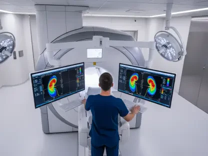
Myocardial perfusion imaging (MPI) plays a pivotal role within nuclear cardiology, being the cornerstone technique for diagnosing critical conditions such as coronary artery disease (CAD) and cardiac amyloidosis. By providing a clear view of blood flow to the heart muscle both at rest and under stress, MPI assists physicians in formulating comprehensive diagnostic and treatment strategies. This non-invasive imaging method has quickly become essential in the management of heart diseases, guiding clinical decisions and improving patient outcomes.
Myocardial Perfusion Imaging (MPI)
Myocardial perfusion imaging (MPI) is a fundamental procedure in nuclear cardiology aimed at evaluating blood flow to the heart muscle. This technique involves taking detailed images of the heart both at rest and during periods of stress, which can be induced through physical exercise or the administration of pharmacological agents. Through this dual approach, physicians can accurately assess the heart’s blood supply and detect any abnormalities that may be indicative of disease.
One of the most significant tools for myocardial blood flow (MBF) quantification in MPI is positron emission tomography (PET). PET is renowned for its precision and ability to deliver highly detailed images and measurements of the heart’s blood flow. Despite its superior quality, PET’s limited availability restricts its widespread use, prompting the exploration of other viable alternatives. Single-photon emission computed tomography (SPECT), although not as precise as PET, is gaining prominence due to its broader accessibility and cost-effectiveness, offering a practical solution to the limitations faced by PET.
PET vs. SPECT in MPI
Positron emission tomography (PET) is the gold standard in myocardial blood flow (MBF) quantification in MPI, known for its unmatched precision and detailed images. The ability of PET to capture the heart’s intricate blood flow dynamics provides clinicians with crucial information necessary for accurate diagnosis and effective treatment planning. However, the deployment of PET is often limited by its availability, requiring sophisticated technology and resources not always accessible in all healthcare settings.
Single-photon emission computed tomography (SPECT) is emerging as a strong contender to PET due to its wider availability and more affordable implementation. Advances in SPECT technology, particularly with the development of cadmium zinc telluride (CZT) cameras, are enhancing the quality and reliability of the images produced. While traditionally considered less precise, these advancements enable SPECT to serve as a robust alternative, facilitating broader access to advanced cardiac imaging and supporting the diagnostic needs of more healthcare facilities.
Non-Invasive Diagnosis of Cardiac Amyloidosis
The shift toward non-invasive diagnostic approaches in nuclear cardiology is exemplified by the preference for nuclear imaging over endomyocardial biopsy (EMB) in diagnosing cardiac amyloidosis. This landmark transition reduces the inherent risks associated with invasive procedures and permits early detection of the disease, which is critical for initiating timely and effective treatment. By leveraging nuclear imaging, clinicians can provide patient care that is not only safer but also more accurate in identifying cardiac amyloidosis.
Critical challenges in diagnosing cardiac amyloidosis include distinguishing between AL amyloidosis and ATTR amyloidosis, both of which require different therapeutic approaches. Accurate differentiation through nuclear imaging is essential for prognosis and customized treatment planning. Advanced imaging techniques provide detailed insights into the heart’s structural and functional changes, thereby enhancing the precision of diagnosis and equipping physicians with the necessary data to devise personalized treatment strategies for their patients.
Radiopharmaceutical Advancements
The field of nuclear cardiology has seen significant advancements with the development of new radiopharmaceuticals, such as the FDA-approved F18-Flurpiridaz. These innovations have considerably expanded the diagnostic toolkit, allowing for more precise and reliable cardiac assessments. Radiopharmaceuticals are vital in enhancing the effectiveness of myocardial perfusion imaging (MPI), providing clearer images and facilitating a more accurate analysis of heart conditions.
The introduction of such advanced radiopharmaceuticals marks a pivotal step forward in the evolution of nuclear cardiology, improving diagnostic accuracy and expanding the capabilities of myocardial imaging. By yielding higher-quality data, these substances facilitate better-informed clinical decisions and improved patient outcomes. The continuous development and approval of new radiopharmaceuticals underscore the field’s commitment to advancing cardiac care through innovation and cutting-edge technology.
Integration of Artificial Intelligence
The integration of artificial intelligence (AI) into nuclear cardiology is poised to revolutionize the field, particularly in the realm of myocardial perfusion imaging (MPI). AI enhances the accuracy and efficiency of quantitative assessments, enabling more precise and timely cardiac diagnostics. By automating complex processes and analyzing large datasets, AI streamlines the evaluation of imaging results, reducing the likelihood of human error and providing clinicians with critical insights more rapidly.
AI’s role in refining cardiac PET-MPI is especially noteworthy, as it enhances the diagnostic capabilities beyond traditional methods. By leveraging machine learning algorithms and advanced data analytics, AI assists in identifying subtle patterns and anomalies that might be missed by conventional analysis. This integration promises to improve diagnostic precision, facilitating more effective treatment planning and patient management by providing healthcare professionals with powerful tools for better decision-making.
Physiological Aspects of Myocardial Blood Flow
Understanding the physiological aspects of myocardial blood flow is crucial for accurate cardiac assessments and effective treatment planning. The heart’s ability to autoregulate blood flow, especially during periods of exercise, is a key consideration in evaluating its health. This autoregulatory mechanism ensures that the heart receives adequate blood supply to meet the increased demands placed upon it during physical exertion.
However, there is a need for further research to fully comprehend the differing physiological responses induced by pharmacological agents versus physical exercise during myocardial perfusion imaging (MPI). These differences can have significant implications for the accuracy and reliability of cardiac assessments. By exploring and understanding these physiological dynamics, researchers can enhance the interpretative accuracy of imaging results, ensuring that diagnostic tools mirror real-life conditions more closely.
Challenges and Future Prospects
Nuclear cardiology has revolutionized how we diagnose and treat cardiovascular issues, offering detailed heart health assessments with minimal invasiveness. This field has made incredible strides, with advanced imaging systems greatly enhancing our ability to detect and manage heart diseases. These advancements lead to higher diagnostic accuracy and more effective treatment planning for patients.
The constant development in imaging techniques and technological progress provides us with increasingly precise and dependable insights into cardiac function. This makes nuclear cardiology an essential tool for cardiologists. Whether using PET scans, SPECT, or other imaging methods, the focus is always on achieving greater precision and reliability.
Moreover, ongoing research and innovations in this field are continually pushing the boundaries of what we understand about heart health, leading to earlier detection of issues that might have gone unnoticed. This results in better patient outcomes, as conditions can be treated more effectively at earlier stages. Overall, the importance of nuclear cardiology in modern medicine cannot be overstated, as it is pivotal in advancing our capabilities to ensure heart health and improve the quality of cardiovascular care.