
In a decisive move that reshapes decades of standard practice, new guidelines from the American Dental Association advocate for a significant evolution in dental imaging, establishing a clear mandate that radiographs should only be ordered based on specific clinical necessity. This updated framework, developed by a panel of experts and published in the January 2026 issue of The Journal of the American Dental Association, represents a foundational shift away from routine, schedule-driven X-rays and toward a more thoughtful, patient-specific diagnostic process. The primary objective is to thoughtfully curtail lifetime radiation exposure for patients and professionals alike, without diminishing the critical role that imaging plays in modern dentistry. This new consensus emphasizes that every decision to use imaging, from traditional two-dimensional X-rays to advanced cone-beam computed tomography (CBCT), must be justified by evidence gathered during a hands-on clinical evaluation, ensuring that technology serves as a targeted tool rather than a standardized procedure for every patient who sits in the dental chair.
The Primacy of the Clinical Examination
The new recommendations firmly establish a thorough clinical examination as the non-negotiable prerequisite to any form of diagnostic imaging, signaling a departure from the long-standing practice of routine radiography. This patient-first philosophy requires dentists to base their decision to order an X-ray on a comprehensive evaluation that includes a detailed review of the patient’s medical and dental histories, an assessment of previously captured images, and, most critically, the direct findings from a physical exam. Dr. Erika Benavides, lead author of the report, drew a compelling parallel to general medicine, explaining that just as a physician wouldn’t order an X-ray without first examining a patient, dental radiographs should follow the same logic. This approach champions a diagnostic model where imaging is used to investigate or confirm specific clinical suspicions, rather than being an automatic component of every visit. The guidelines, which have been formally endorsed by the American Academy of Oral and Maxillofacial Radiology (AAOMR), underscore a broad consensus within the dental community to prioritize individualized care and justified radiation use.
This fundamental shift empowers clinicians to tailor diagnostic pathways to each patient’s unique health profile, moving beyond a one-size-fits-all methodology. By mandating a clinical exam as the initial step, the guidelines encourage a more investigative and evidence-based practice. Instead of relying on a predetermined schedule, practitioners are now directed to use their professional judgment to determine if imaging is truly necessary to answer a specific diagnostic question that cannot be resolved through other means. This method not only enhances the precision of diagnoses but also strengthens the patient-dentist relationship by fostering transparent conversations about the rationale behind ordering a particular test. This deliberate and justified approach ensures that the benefits of obtaining a radiograph—such as detecting hidden decay or assessing bone levels—distinctly outweigh the minimal but cumulative risks associated with radiation exposure, aligning dental care more closely with the broader principles of modern, personalized medicine and patient safety.
Tailored Guidance for Common Conditions
A significant strength of the updated recommendations lies in the detailed guidance provided for a wide array of common clinical scenarios, equipping dentists with a practical framework for making informed decisions. When it comes to the detection of dental caries, the paper avoids broad-stroke rules, instead offering nuanced advice for different tooth surfaces, including anterior proximal, posterior proximal, occlusal, and root surfaces. It reinforces that the selection between various radiographic techniques, such as bitewing or periapical views, must be dictated by the dentist’s professional clinical judgment. This decision should consider critical factors like the suspected location of a lesion and the patient’s specific anatomy—for instance, whether the spaces between teeth are open or closed, which can impact the visibility of interproximal decay. This level of specificity encourages a more analytical approach, ensuring that the chosen imaging modality is the most effective one for the particular diagnostic challenge at hand, thereby maximizing the diagnostic yield while minimizing unnecessary exposure.
In the complex field of periodontal disease management, the guidelines reaffirm the essential role of radiographic evaluation for making an initial diagnosis and establishing a crucial baseline for monitoring disease progression and the effectiveness of treatment. However, the document explicitly pushes back against fixed imaging schedules for follow-up care. Instead, it states that the frequency of subsequent radiographic examinations should be determined dynamically, based on ongoing clinical findings and how the patient is responding to therapy. The recommendations uphold the combination of a 2D full-mouth radiographic series paired with a comprehensive clinical examination as the current gold standard for the thorough evaluation of periodontal disease. Importantly, the panel concluded that there is currently no evidence to support the routine use of CBCT for the general management of periodontitis, reserving its powerful three-dimensional imaging capabilities for the treatment planning of particularly complex cases where its detailed view is truly indispensable for a successful outcome.
Specialized Applications and Patient Profiles
Further reinforcing the commitment to personalized care, the guidelines present a structured set of recommendations tailored to different types of dental appointments and patient profiles. A clear distinction is made between the imaging needs of a new patient undergoing an initial comprehensive evaluation and a returning patient attending a routine recall visit. This guidance is further stratified by key demographic and health factors, including the patient’s age, their current stage of dental development, and their individual risk factors for prevalent conditions such as dental caries and periodontal disease. For example, a young adult with a history of high caries activity and closed contacts between their teeth would have a different imaging prescription than a middle-aged patient with excellent oral hygiene and no new clinical findings. This multi-layered approach ensures that imaging protocols are not arbitrary but are instead meticulously customized to the specific circumstances of each individual, promoting both diagnostic accuracy and patient safety.
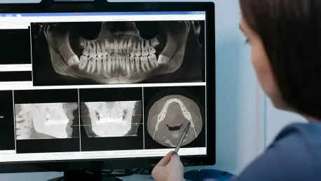
The document also extends its detailed guidance to a variety of dental specialties, outlining best practices for the use of imaging in specific and often complex clinical contexts. For oral and maxillofacial surgery, panoramic radiography is recommended for the initial assessment and treatment planning related to third molars and other anomalous teeth. In orthodontics, panoramic radiographs are identified as the preferred initial tool for monitoring tooth eruption patterns and evaluating root alignment during treatment. For implant dentistry, a two-tiered strategy is advised, beginning with panoramic radiography for the initial site assessment, followed by the more detailed CBCT for precise presurgical planning. In endodontics, a clear hierarchy is established, with intraoral 2D radiographs serving as the primary modality and limited-field CBCT designated as a secondary tool for complex cases. A special emphasis is also placed on the judicious use of radiographs for pediatric patients, mandating effective dose-reduction techniques to protect them during critical years of development.
A Culmination of Safety and Best Practices
This comprehensive publication on clinical necessity is the second installment of a two-part series from the ADA aimed at thoroughly optimizing the use of diagnostic imaging in dentistry. The first set of recommendations, which were released in 2024, focused squarely on the critical issues of radiation safety and evolving regulatory matters. One of the landmark findings from that initial paper was the formal recommendation to discontinue the routine use of both thyroid and abdominal shielding during dental imaging procedures. This progressive stance built upon earlier guidance from the AAOMR and reflected a growing body of evidence indicating that modern, well-collimated dental X-ray equipment makes such shielding redundant and, in some cases, potentially obstructive to obtaining a clear diagnostic image. Together, these two reports form a cohesive and modern framework designed to guide dental professionals toward safer, more effective, and more justified imaging practices for all patients.
Ultimately, the new clinical guidelines were anchored in the foundational radiation safety principle of ALARA—maintaining exposure “As Low As Reasonably Achievable.” Dr. Benavides provided reassurance by noting that dental X-rays are generally very safe, sometimes delivering a radiation dose lower than what a person receives from a single day of natural background exposure. Despite this high safety profile, the core message delivered by the ADA was one of conservative and thoughtful application. The ADA and AAOMR urged all dental professionals to thoroughly review and implement these evidence-based recommendations in their daily practice. This call to action fostered a culture of mindful imaging, encouraging open and transparent communication with patients about both the significant benefits and the minimal risks associated with any radiographic examination, thereby ensuring that every X-ray taken was a necessary one.