
The precise and early diagnosis of prostate cancer remains a significant challenge in modern medicine, as current methods like serum PSA testing often lack the specificity needed to avoid over-diagnosis and subsequent unnecessary, invasive biopsies. Conventional ultrasound imaging, while widely used, struggles to differentiate between benign and malignant lesions and cannot visualize key molecular indicators like prostate-specific membrane antigen (PSMA) in real-time. This diagnostic gap has fueled the search for more sophisticated technologies capable of detecting cancer at its molecular roots, paving the way for ultrasound molecular imaging. This advanced approach uses targeted probes to illuminate specific biomarkers, offering the potential for a more accurate and timely diagnosis. However, the advancement of this promising field has been persistently hindered by the cumbersome and inefficient methods used to create these essential probes, a bottleneck that has limited their widespread clinical application and spurred researchers to seek a completely new manufacturing paradigm.
A Biological Blueprint for Advanced Imaging
Ultrasound molecular imaging technology represents a promising frontier for the early detection of prostate cancer by enabling the visualization of PSMA expression at the molecular level. Unlike conventional imaging, this technique relies on specialized probes designed to bind to specific cancer biomarkers. The conventional method for creating these probes is a multi-step chemical synthesis process that is both complex and fraught with inconsistencies. This strategy typically involves activating a targeting ligand, purifying it, and then covalently attaching it to microbubbles using crosslinkers. The entire procedure can take three to five days and suffers from significant drawbacks, including random orientation of the targeting ligands, which can reduce their effectiveness, and considerable batch-to-batch heterogeneity, making standardized production difficult. Furthermore, the chemical crosslinkers used in this process can trigger immunogenic reactions, raising safety concerns for their potential use in clinical settings. These limitations highlight an urgent need for a more streamlined, reliable, and safer method for producing targeted imaging agents.
A transformative solution to these challenges has emerged from the microbial world in the form of gas vesicles (GVs), which are evolutionarily conserved, gas-filled organelles originally discovered in certain bacteria and archaea. These nanostructures, typically spindle- or cylinder-shaped, are composed of a pure protein shell that is uniquely suited for ultrasound applications. The thin but strong shell has a hydrophobic inner surface that allows gas to diffuse in and out while repelling liquid water, creating a stable gas compartment. This structure allows the GVs to collapse and reform under specific acoustic pressures, generating the nonlinear ultrasound signals essential for high-contrast imaging. Crucially, the assembly of these vesicles is governed by a cluster of genes. One of these, GvpC, codes for a scaffolding protein that strengthens the vesicle and features a C-terminal domain that can be genetically fused with other functional proteins. This provides a natural, programmable scaffold for designing targeted probes that can specifically bind to biomarkers like PSMA, offering a biological alternative to chemical synthesis.
Engineering a Novel Biosynthesis Platform
To overcome the challenges associated with previous genetic engineering attempts, such as steric hindrance and imbalanced protein expression, researchers have developed a sophisticated dual-plasmid thermal induction system within E. coli. This innovative approach separates the production of the gas vesicle scaffold from the synthesis of the targeting component. First, a DNA fragment containing the GV gene cluster from Serratia sp. ATCC39006, but with the GvpC gene removed, was inserted into a pET-28a vector. This created the pET-28a-ΔGvpC-eGVs plasmid, which directs the robust formation of the basic GV structure. Concurrently, a high-affinity PSMA nanobody gene was fused to the C-terminus of the GvpC gene and cloned into a separate, temperature-sensitive pBV220 plasmid. This second plasmid, pBV220-PSMA-GvpC, is responsible for producing the targeting fusion protein. By co-transforming both plasmids into E. coli, an engineered strain capable of producing the complete, targeted probe was created.
The assembly of the final probe, termed PSMA-eGVs, is controlled through a precise, two-step thermal induction program that ensures proper folding and integration of all components. In the first step, the engineered E. coli are cultured at 30°C and treated with an inducer molecule (IPTG), which triggers the expression of the genes on the pET-28a-ΔGvpC-eGVs plasmid to build the foundational GV scaffolds. Once these structures have formed, the culture temperature is raised to 42°C. This thermal upshift activates the promoter on the pBV220-PSMA-GvpC plasmid, initiating the expression of the GvpC-PSMA fusion protein. The N-terminal domain of the newly synthesized GvpC protein then naturally recognizes and binds to the surface of the pre-formed GV scaffolds, effectively decorating them with the PSMA-targeting nanobody. This controlled, sequential assembly process avoids the protein misfolding and stoichiometric imbalances seen in single-plasmid systems, resulting in the efficient and reliable biosynthesis of functional, targeted nanoprobes ready for purification and subsequent use in diagnostic imaging.
Characterization and Validation of the Probes
Following the successful biosynthesis, the resulting PSMA-eGVs and a control version expressing a green fluorescent protein (EGFP-eGVs) were rigorously characterized to confirm their structural integrity and functional capabilities. Transmission electron microscopy revealed that both types of engineered vesicles possessed the typical cylindrical structure with a uniform morphology, consistent with naturally occurring gas vesicles. Further analysis determined the average particle size of PSMA-eGVs to be approximately 125.90 nm with a negative surface potential of -27.28 mV, nearly identical to the EGFP-eGVs. This uniformity is critical for predictable behavior in a biological system. Stability assessments conducted over a 30-day period demonstrated that the PSMA-eGVs maintained a consistent size distribution and surface charge, confirming their structural robustness and suitability for storage and future use.
The functional performance of the engineered probes was first evaluated in vitro to assess their ultrasound imaging capabilities. When suspended in an agarose phantom at various concentrations, both PSMA-eGVs and EGFP-eGVs produced a strong, concentration-dependent enhancement of the ultrasound signal. The signal intensities were comparable between the two probe types at equivalent concentrations, indicating that the addition of the PSMA-targeting nanobody did not interfere with the acoustic properties of the gas vesicles. Furthermore, proteomic analysis using mass spectrometry confirmed the successful construction of the probes. The analysis showed a complete absence of the GvpC protein in the knockout scaffold vesicles, while its abundance in the final EGFP-eGVs and PSMA-eGVs was comparable to that of wild-type vesicles. This comprehensive characterization collectively validated the structural and functional reliability of the genetically engineered probes, clearing the way for testing their targeting specificity.
Demonstrating Specificity in Cellular and Animal Models
The crucial capability of a molecular imaging agent is its ability to specifically target its intended biomarker. In vitro studies were conducted to systematically evaluate this property. Using flow cytometry, FITC-labeled PSMA-eGVs were shown to bind specifically to LNCap prostate cancer cells, which express high levels of PSMA. In contrast, the control EGFP-eGVs exhibited minimal binding. This targeting was confirmed to be PSMA-mediated, as pre-blocking the PSMA receptors on the LNCap cells with a free anti-PSMA antibody significantly reduced the binding efficiency of the probes. As expected, neither probe showed significant binding to PSMA-negative PC-3 cells. Quantitative analysis revealed that 81% of LNCap cells showed fluorescence enhancement after treatment with PSMA-eGVs, compared to only 7% with the control probes. These findings were corroborated by cell adhesion assays, which visually demonstrated substantial binding of PSMA-eGVs to LNCap cell monolayers, a result that was again absent in control groups and PSMA-negative cells.
Building on these promising in vitro results, the imaging performance of PSMA-eGVs was evaluated in tumor-bearing mouse models. Mice with either PSMA-positive LNCap tumors or PSMA-negative PC-3 tumors received systemic injections of the probes. In the PSMA-negative PC-3 tumors, the ultrasound signal from both PSMA-eGVs and control EGFP-eGVs enhanced rapidly and then decayed quickly, indicating no specific retention. Conversely, in PSMA-positive LNCap tumors, the PSMA-eGVs demonstrated significantly prolonged signal retention compared to the control probes. While initial peak signals were similar, at three minutes post-injection, the signal from PSMA-eGVs remained strong, whereas the signal from EGFP-eGVs had substantially faded. By ten minutes, the targeted probes generated an intensity 8.5 times higher than the controls. This sustained signal enhancement confirms that the PSMA-eGVs actively bind to and accumulate within PSMA-expressing tumors in a live animal model, demonstrating their efficacy for in vivo targeted imaging.
Correlating Biosynthetic Probes With Clinical Standards
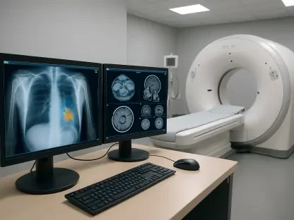
To validate the diagnostic relevance of the ultrasound signals generated by the PSMA-eGVs, their performance was compared against established clinical benchmarks: positron emission tomography/computed tomography (PET/CT) imaging and immunohistochemical (IHC) tissue analysis. In the same tumor models, PET/CT scans using the clinical tracer 18F-PSMA revealed substantial radiotracer uptake in LNCap tumors, confirming high PSMA metabolic activity, while no significant uptake was observed in PC-3 tumors. Similarly, IHC staining of excised tumor tissues showed clear and abundant PSMA protein expression in LNCap cells, which was absent in PC-3 cells. These results from gold-standard clinical methods provided a definitive map of PSMA expression against which the novel ultrasound probe could be measured.
A linear regression analysis was then performed to correlate the data from all three modalities. The results demonstrated a remarkably strong positive correlation between the ultrasound signal intensity (measured as area under the curve) from PSMA-eGVs and both the PET/CT signal (SUVmax) and the PSMA expression levels quantified by IHC. In PSMA-positive LNCap tumors, the correlation coefficient (R²) between the UMI signal and PET-CT SUVmax was 0.90 for small tumors and 0.94 for large tumors. Similarly, the correlation with IHC-quantified PSMA expression yielded an R² of 0.83 for small tumors and 0.98 for large tumors. These strong correlations prove that the biosynthetic probes can accurately and quantitatively reflect the underlying levels of PSMA expression in tumors. This finding suggests that ultrasound molecular imaging with PSMA-eGVs has the potential to provide diagnostic information comparable to PET-CT but with the advantages of being non-invasive, radiation-free, and more cost-effective.
A Promising Path to Clinical Translation
This investigation successfully developed and validated a one-step biosynthesis platform for the efficient production of targeted ultrasound molecular imaging probes. Using PSMA-positive prostate cancer as a proof-of-concept model, the study demonstrated that the biosynthesized PSMA-eGVs exhibited excellent physical stability and high targeting specificity both in vitro and in vivo. The strong correlation established between the ultrasound imaging signals and established diagnostic standards like PET-CT and immunohistochemistry underscored the probe’s ability to accurately reflect tumor biomarker expression. This work provided a promising strategy for developing targeted ultrasound probes for the non-invasive diagnosis of tumors, potentially offering a safer, more accessible, and lower-cost alternative to current methods. The streamlined and adaptable nature of the genetic engineering platform suggested it could be readily modified to produce probes for a wide range of other diseases and molecular targets, opening new avenues for diagnostic imaging.