
Surgeons are often lauded for their steady hands and expertise. Life-saving surgeries require not just advanced medical skills and knowledge, but tools as well. In the ever-evolving world of medical technology, precision is key.
One of the most critical tools in their arsenal is the endoscope, an essential device for diagnosing and treating a variety of conditions. However, traditional endoscopes are not without limitations, particularly when it comes to accessing smaller, more intricate areas of the body.
Enter a groundbreaking development from the University of Washington: a new metalens technology designed to improve both the size and functionality of endoscopes, offering a clearer, more precise view of the body’s internal systems.
Here’s a closer look at what the research team has uncovered.
The Evolution of the Endoscope
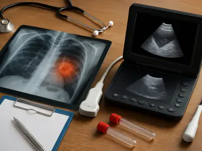
The endoscope is an invaluable medical device used and adapted for a number of simple procedures. Endoscopes are flexible, long tubes with a camera on one end and a lens on another. The lens end is inserted into the patient, with light provided by optical fibers, and the video camera projects and magnified view on a screen.
What makes endoscopes effective is their structure; they are long and winding, yet small in diameter. Endoscopes are critical for the detection, viewing, and treatment of several diseases and are capable of finding blood clots in the heart and airway blockages.
Endoscopes are also the tool of choice for performing biopsies (removal of small tissue for testing), which are key in diagnosing cancer.
Despite its widespread and essential use, the endoscope has its limitations. There are a number of unmet medical needs which endoscopes cannot be used for, due to their size. Specialists need a device that can access smaller parts of the body, like arteries and bronchioles. The other prevailing issue lies with the camera lens and light source, which are critical to viewing diseased and damaged tissue.
Working to solve these issues is a research team at the University of Washington’s Department of Electrical and Computer Engineering and Physics. Under the leadership of Professor Arka Majumdar, the team has focused on developing a new lens system that will give physicians an improved view of intricate areas in the body.
While metalenses are not a new technology, applying this technology to endoscopes is a novel application. Unlike normal lenses—which are curved—metalenses are flat with tiny surfaces (nanostructures) that manipulate light. Professor Majumdar and his team were able to engineer an endoscope that makes use of metalens technology to improve internal visibility in the body and access smaller spaces. Their findings were published in Light: Science & Applications.
Professor Majumdar evaluated his team’s success, stating that “A lot of people developing endoscopy are working on the software, the computational side. Our team chose to develop a metalens for the optical hardware in an endoscope, and as our work progressed, we realized that many improvements were possible.”
How the Metalens Improve the Endoscope
One of the key benefits to using a metalens in an endoscope is its dimensions. The University of Washington research team was able to create a metalens so small that it would reduce the size of the smallest endoscope by half. For physicians, the possibilities are endless. With smaller medical devices, they’ll be able to access parts of the body that have never been seen before with optical imaging.
This technology will have a major impact on patients at risk for cardiac arrest and stroke. With smaller, more nimble endoscopes and greater visibility, physicians will be able to assess and access blood clots in the brain and diseased arteries in the body.
Physicians have a number of benefits to look forward to once the metalens endoscope is commercially available:
Real-time visual feedback
Improved efficiency
Reduced medical errors
Increased procedure success rate
Enhanced visuals when compared to X-rays
Eric Seibel, a co-author and UW research professor, explains, “We are trying to extend the eyes of the surgeon or the physician deeper into the body […] This is an area that I have been working in for 25 years, and this new technology has the potential to leapfrog over my past work. I’m very excited about this device.”
Leveraging chromatic aberration in a metalens
A special technique known as quantitative phase imaging is used to enhance the endoscope using a metalens. Simply put, these tiny lenses adopt a microscope-effect, by measuring light waves as they pass through the transparent lens. Using depth sensors, a 3D render is produced in full-color and is seen as a “video”. The simple elegance of this system lies in the fact that very little computational power is required. This small tool, measuring at a width of 0.5mm, has powerful applications and implications.
While quantitative phase imaging is by no means a new field of study, the application in an endoscope is novel. This is what has captured attention in the industry and generated eager anticipation for the prototype.
The research team has also been able to turn a problem into a solution. Chromatic aberration is the term used to describe a colored fringe that appears on the edge of an image due to the fact that light hasn’t been concentrated into a focal point. This is widely considered an issue that must be addressed, but the UW team has found a purpose for this.
According to Aamod Shankar, the lead author of the research paper, they have been able to “create a chromatic splitting along focus, causing each color to converge at a different depth. In the reverse direction, the longitudinal rainbow effect allows for mapping depth into the color channels of a camera.”
Essentially, chromatic aberration is used as a tool for depth sensing.
A vision for the future
The research team at the University of Washington will dedicate the next two years to developing a prototype. Metalenses are a well-researched field of study and development, which strengthens the bid to develop a market-ready product. However, while there is a strong case for metalens endoscopes, it will be at least a decade before physicians can expect to acquire this medical device.
“This chromatic aberration providing information about depth is good, but this is still experimental,” Majumdar said. “To get what a surgeon needs, we need to improve the image quality, and that’s what we are pushing toward.”
There’s no doubt, however, about the impact a smaller, more powerful endoscope will have on the field of medicine. Diagnostic capabilities will increase, while invasive surgeries are likely to decrease.
Concluding Thoughts
Researchers at the University of Washington imagine a new view for physicians by creating a proof of concept for a metalens endoscope. This is a medical device that is instrumental to the detection, diagnosis, and treatment of various diseases.
Looking to resolve some of the limitations of this device, they’ve incorporated metalens technology, which not only improves medical imaging, but has the capability to reduce the size of endoscopes by almost 50%.
With smaller endoscopes that can provide better visibility, medical practitioners will be able to see into smaller spaces of the body. This will be a major breakthrough for patients at risk of cardiac arrest or strokes, as they’ll be able to detect clogs and blood clots in smaller spaces like arteries and bronchioles in the lungs.
This breakthrough combines chromatic aberration—a typically undesirable optical effect—with quantitative phase imaging to create high-resolution, real-time 3D imaging. The research team aims to refine image quality and produce a prototype for clinical testing within two years.
If successful, this innovation could significantly enhance the precision and safety of medical procedures, reducing errors and improving patient outcomes, particularly in treating cardiovascular diseases. While widespread adoption may take decades, the technology promises to revolutionize endoscopic applications and healthcare.