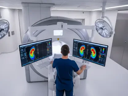

Medical professionals frequently encounter a significant bottleneck when attempting to integrate high-resolution point-of-care ultrasound into daily practice due to the intensive training required to master anatomical interpretation. While the hardware itself has become increasingly portable and affordable, the ability to discern nuanced structures within “grainy” grayscale images often remains limited to specialists with years of experience. Clarius Mobile Health has addressed this specific barrier through the release of the Clarius App 12.2.6 update, which introduces the specialized T-Mode Heart and T-Mode Knee AI-powered tools. These additions are designed to act as a real-time bridge between raw imaging data and clinical decision-making by utilizing artificial intelligence to clarify anatomical landmarks. By providing an educational overlay directly onto the live feed, the system allows non-specialists to perform diagnostic screenings with a level of confidence that was previously difficult to achieve without external consultation or extensive postgraduate certification.
Bridging the Gap in Diagnostic Imaging
Overcoming the Interpretation Barrier in Modern Medicine
The primary challenge in the widespread adoption of point-of-care ultrasound is the “interpretation gap,” where a clinician can physically capture an image but struggles to identify the specific pathology within complex structures. For decades, interpreting the rapid movements of heart valves or the subtle layering of knee ligaments required a highly specialized eye trained to see past the inherent noise of ultrasound waves. T-Mode technology effectively solves this by providing a dual-view interface where the traditional grayscale image is paired with a synchronized, AI-generated replica. This replica uses distinct color-coding and labels to highlight essential anatomical markers, essentially providing a digital textbook overlay in a live clinical setting. This immediate visual feedback helps the practitioner confirm that they are looking at the correct structure, such as the mitral valve or the patellar tendon, thereby reducing the ambiguity that often leads to diagnostic errors or unnecessary specialist referrals in busy primary care environments.
Furthermore, the integration of AI-driven guidance serves as a continuous learning tool that empowers general practitioners to expand their diagnostic scope without leaving their clinics. When a user activates T-Mode during a cardiac scan, the software identifies and tracks the heart’s chambers in real-time, assisting the learner in understanding spatial relationships that are often confusing on a standard monitor. This capability is not just about identifying anatomy; it is about providing the user with the certainty needed to make a quick clinical call. By simplifying the visual output, Clarius has successfully lowered the technical threshold for entry, making it possible for emergency physicians and primary care doctors to utilize ultrasound as a standard diagnostic tool rather than a specialized elective. This shift ensures that the technology is utilized to its full potential, rather than sitting idle due to a lack of confidence in interpretation, which ultimately benefits the patient through more immediate and accurate bedside assessments.
Enhancing Clinical Efficiency and Frontline Care
The strategic implementation of T-Mode Heart significantly alters the workflow for screening common yet serious conditions like heart failure and valvular disease during routine examinations. In traditional settings, a patient presenting with vague symptoms such as chronic fatigue or shortness of breath might wait weeks for an echocardiogram at a specialized imaging center. With these new AI tools, a primary care physician can perform a preliminary cardiac sweep and use the color-coded labels to verify chamber size and function instantly. This streamlined approach allows for the early detection of abnormalities, ensuring that high-risk patients are prioritized for advanced care while those with healthy cardiac function are spared unnecessary anxiety and expense. The ability to distinguish between different cardiac structures with AI assistance transforms the ultrasound from a passive recording device into an active diagnostic partner that supports the clinician’s expertise at every step of the process.
In the realm of musculoskeletal health, the T-Mode Knee tool provides a similar efficiency boost for clinicians managing one of the most common complaints in general practice. Knee pain often requires a complex differential diagnosis to determine whether a patient needs conservative physical therapy or an urgent orthopedic surgical consultation. The AI tool assists the clinician in navigating all planes of the joint, ensuring that specific structures like the meniscus or the collateral ligaments are properly visualized and assessed. By providing a clear, labeled view of the knee’s internal geometry, the technology helps the provider make an informed decision on the spot. This democratization of high-level imaging means that the frontline of healthcare becomes more self-sufficient, reducing the overall burden on the healthcare system and allowing specialists to focus on the complex cases that truly require their expertise.
Improving Patient Outcomes Through Applied AI
The deployment of these automated interpretation tools established a new standard for how handheld medical devices can support clinical workflows without requiring significant overhead. Clinicians moved toward a model where diagnostic imaging occurred at the first point of contact, which effectively minimized the diagnostic delays that previously hindered patient recovery. For future implementations, healthcare organizations should consider integrating these AI-guided platforms into standard residency training programs to accelerate the proficiency of the next generation of physicians. Furthermore, stakeholders should focus on expanding similar AI overlays to other complex anatomical regions, such as the abdominal organs or the carotid arteries, to create a comprehensive diagnostic ecosystem. This evolution will likely lead to a healthcare environment where the initial physical exam is augmented by immediate internal visualization, resulting in a more proactive and precise approach to population health management.